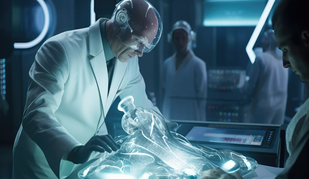
Understanding Nuclear Medicine Procedures

When you get an x-ray or CT scan at the hospital, these tests use forms of radiation to see inside your body and make pictures of your organs and bones. As per experts in the field like PRP Imaging, nuclear medicine is a branch of medicine that also uses radiation, but in a special way to not just image, but diagnose and treat disease. This article will explain what nuclear medicine is, some types of nuclear imaging tests, and why Nuclear Medicine Procedures are so useful for patients.
What is Nuclear Medicine?
Nuclear medicine uses small amounts of radioactive material, called radiotracers or radiopharmaceuticals, that are typically injected into the bloodstream, swallowed, or inhaled as gases. The radiotracer travels through the area being examined, giving off energy. Special cameras detect this energy and create pictures of the body. Unlike x-rays though, nuclear imaging lets doctors see how well organs and tissues are working, not just their structure.
Nuclear Tests to Diagnose Problems
Here are some common types of nuclear imaging tests:
PET Scan - Positron emission tomography (PET) scan uses radiotracers containing positrons. Positrons react with electrons in the body, emitting energy that the PET scanner detects. PET scans show abnormal activity, like cancer, inflammation, heart disease or brain disorders.
SPECT Scan – Single photon emission computed tomography (SPECT) is similar to PET but uses gamma ray emitting radiotracers. SPECT also produces 3D images of organs and tissues and blood flow to detect problems.
Thyroid Scan – For thyroid scans, patients swallow a radioactive iodine solution which is taken up by thyroid cells. The scan images this activity to identify thyroid problems and tumors.
Bone Scan – Bone scanning radiotracers like technetium-99 concentrate in areas of bone remodeling. A scanner creates an image highlighting problems like fractures, arthritis, or cancer.
Cardiac Stress Test – Patients exercise on a treadmill and are injected with a radiotracer while their heart is pumping hard. Scans show blood flow during stress, diagnosing heart disease.
The unique ability of radioactive materials to concentrate in certain tissues allows doctors to pinpoint medical conditions that other tests might miss. The imaging shows not just anatomy, but actual physiology and function.
Using Nuclear Medicine to Treat Disease
Besides diagnosis, nuclear medicine can also treat some conditions. Two examples are:
Radioactive Iodine (RAI) – RAI is used to destroy overactive thyroid tissue and thyroid cancer cells without surgery. Patients swallow a capsule or liquid, and the radioactive iodine kills the abnormal cells.
Radiation Therapy – Small sealed radioactive implants placed inside or near a tumor deliver targeted radiation to kill cancer cells. The implants may stay for minutes or days.
RAI and radiation therapy allow powerful treatment that harms cancer but spares healthy tissue. The radioactive material is carefully controlled to provide healing benefits.
The Value of Nuclear Medicine
Nuclear medicine has many advantages:
- Finds disease in early stages before anatomy changes
- Screens the entire body with only one scan
- Identifies whether tumors are benign or malignant
- Guides doctors about proper treatments
- Avoids exploratory surgery by confirming or ruling out disease
- Treats certain conditions non-surgically
Conclusion
The unique views into body function make nuclear imaging extremely useful. It provides information other tests cannot. Nuclear medicine helps doctors diagnose illness as soon as possible and select the best therapy. For many patients, nuclear medicine plays a lifesaving role.


